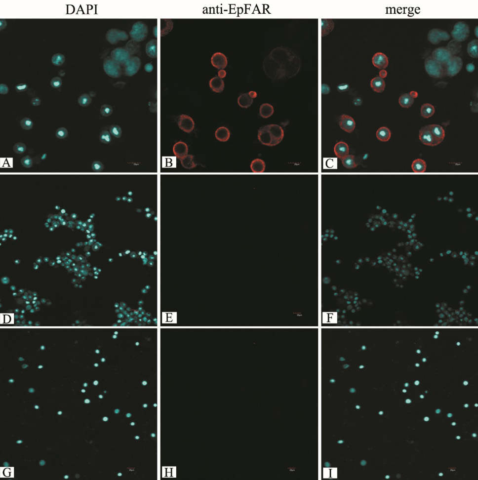
Fig. 1.
Localization of EpFAR in Sf9 cells. Sf9 cells were infected with the recombinant baculovirus and fluorescence imaged by LSCM. A, B, and C, cells were infected with the recombinant nuclear polyhedrosis baculovirus including EpFAR. D, E, and F, cells treated with the virus generated with an empty pFastBac HT B vector were used as a control. G, H, and I, cells cultured in the normal culture medium were used as another control.